

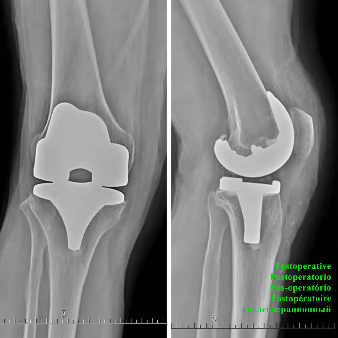
Knee System Get Back Living Life Without Knee Pain
Knee Joint: the knee joint is a compound joint. It is composed of the medial and lateral condyles of the femur and the medial and lateral tibia, as well as the patella, ligaments, muscles, etc. It is the joint with the largest and most complex structure in the human body and more chances of injury.
Artificial implants made to relieve knee joint pain, restore knee joint structure and function, and correct joint deformities.
Surgical Indications:
Severe joint pain, instability, deformity, severe disturbance of activities of daily living, cases of ineffective or unsatisfactory results after conservative treatment:
1: Various inflammatory arthritis and osteoarthritis of the knee joint, rheumatoid arthritis, hemophilic arthritis; 2: A small number of severe traumatic arthritis; 3: Osteoarthritis after failure of high femoral osteotomy Inflammation; 4: Patellar osteoarthritis in a small number of elderly people; 5: Infectious arthritis has been cured or resting; 6: A small number of primary or secondary osteochondral necrotic diseases.
 pelvis
femoral head
femur
pelvis
femoral head
femur
1) Open Femur Pulp
2) Assemble Instruments
3) Set Valgus Angle
4) Adjust Osteotomy Volume
5) Fix Osteotomy Template
6) Osteotomy
Important Hints:
1. The thickness of the femoral condyle products for straightening and knee flexion is 9mm.
2. The connector can be connected and disconnected only when the knob is in the unlocked position.
3. When punching the fixing nails, punch the 0 position first, and then punch the oblique hole after the final confirmation.
4. If the headed screw is selected when fixing the osteotomy template, it should not be screwed too tightly to cause excessive pressure.

Acetabular Component
Femoral Head
Acetabular Liner
Femoral Stem
1) Assemble the tool
2) Adjust the force line
3) Adjust the back inclination angle
5) Adjust the amount of osteotomy
6) Fix osteotomy template
7) Osteotomy
8) Inspection of clearance and force line
Important Hint:
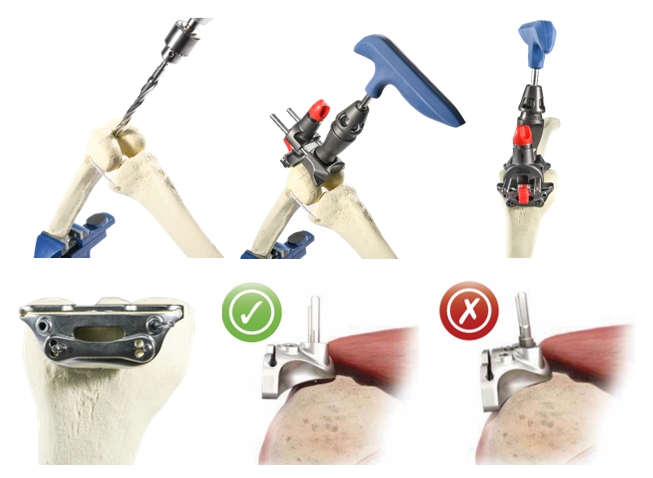
1. The anterior long slot is used to insert temporary nails during the operation to limit the left and right movement of the osteotomy template;
2. The minimum thickness of tibial tray + meniscus is 10mm.
3. When the standard insert of the tibial osteotomy measuring instrument is inserted into the saw blade slot, the adjustment range of the osteotomy volume is 0-14mm, and when the +4 insert is inserted into the saw blade slot, the adjustment range of the osteotomy volume is 4-18mm.
Important Hint:
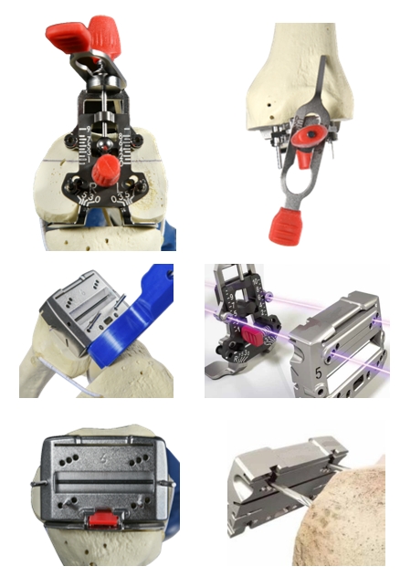
A. After the femoral condyle measurer hugs the posterior condyle, fixation pins can be used to temporarily fix the measurer;
B. The red knob of the femoral condyle can be easily adjusted by pressing the red knob hard, and the loosening resistance will increase, which is convenient for positioning
C. The front refers to the upper hole for surgery, and the rear refers to the lower hole for surgery;
D. The four-in-one osteotomy template can be adjusted back and forth by 1.5mm to balance the amount of osteotomy; its 0-position guide groove can help guide the installation.
E. When fixing the four-in-one bone plate, it is more secure to use threaded headed screws when the two oblique holes are fixed (especially for patients with osteoporosis). Side bevel nails.
F. The advantage of using a combined four-in-one posterior condylar osteotomy is that it is convenient to measure the knee flexion gap before the formal osteotomy.
G. After removing the osteotomy template, carefully handle the uncut residual bone along the osteotomy surface with an osteotomy knife or an oscillating saw.
General Nursing
Observation of incision drainage tube: due to the use of tourniquets during the operation of knee arthroplasty, vascular reactive dilation and bleeding of vascular stump at the site of intra-articular tissue resection are often caused after surgery. Blood infiltration of incision dressing and color, quality and quantity of drainage should be closely observed. The catheter was extubated 24-48 hours after the postoperative drainage, when the drainage volume was less than 20ml/d. To keep the drainage tube in the process of drainage unobstructed, prevent distortion, folding and congestion, once every 30 minutes extrusion, such as found fast drainage tube flow velocity is higher than 100 ml/h, shall notify competent doctor, to keep the incision dressing clean and dry, once the pollution changes in time, the orders timely and correct use of antibiotics gauze, preventing incision infection.
Postoperative position: After operation, patients should take off the pillow and lie supine for 6 hours. After 6 hours, patients should take supine position. The soft pillow behind the knee of affected limb should be raised to maintain neutral position, so as to avoid excessive compression of the calf gastrointestinal muscle and the common nerve, resulting in thrombosis of the calf gastrointestinal venous plexus and injury of the common nerve.
Observation of blood supply of affected limbs: Pay close attention to the sensation of affected limbs, skin temperature of affected limbs, skin color and pulsation of dorsal pedal artery, and deal with abnormal cases in time.
Postoperative Complications
Postoperative Infection
Including wound and intra-articular infection, urinary tract infection, pulmonary infection and so on, can lead to fever. Therefore, we should closely observe the changes of patient's body temperature and blood liquefaction test value, keep the wound dressing dry, and replace it in time after contamination. Wound infection mostly occurs in 1-3 weeks after operation. The patient complains that the pain is aggravated or relieved and then aggravated, the body temperature is increased, the pulse is fast, the leucocyte is increased, and there is local redness, swelling, fever and pain.
Prevention: 1. Nursing of the urinary catheter: It is better to keep the urinary catheter for 24 hours and keep it unobstructed. Perineal care should be done daily to encourage patients to drink more water. 2. In order to prevent pulmonary infection, teach patients to breathe deeply and cough effectively, cough up phlegm as much as possible, kowtow on the back of patients in the morning and evening nursing every day, and inhale atomized whenever necessary. 3. Pressure sore prevention: due to poor blood circulation of the elderly, long-term bed rest after surgery, less activity, low body reaction, pressure sore is easy to occur, should increase nutrition, keep patients’ sheets dry and clear, avoid body and bed contact friction and shear force, reduce the pressure on tissues. When lying flat, the patient's toes are pointed upward, and sponge pads can be padded under his feet to prevent pressure sores on his heels. Regular help patients turn over, can reduce the formation of back pressure sores.
